Key takeaways
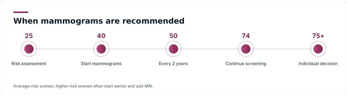
- The U.S. Preventive Services Task Force now recommends biennial mammography for average-risk women beginning at age 40 and continuing through 74.
- Other major organisations recommend annual screening from 40 or 45. The disagreement is about interval and starting age, not about whether screening works.
- Since 2024, U.S. facilities must tell patients whether their breasts are dense, because dense tissue both raises risk and hides cancers on mammography.
- Women at high risk — including those with BRCA variants, a strong family history, or prior chest radiation — need earlier screening and usually annual MRI.
- False positives and callbacks are common and are the main downside of screening. They are not evidence the test failed.
Breast cancer is the most commonly diagnosed cancer in American women, with roughly one in eight developing it over a lifetime. It is also the screening topic where reasonable experts most publicly disagree — not about whether mammography saves lives, which the evidence supports, but about when to start, how often to repeat, and how to weigh the harms of finding things that would never have caused trouble. Here is what changed, what is settled, and how to build a plan.
What changed in the guidelines
For years, the USPSTF recommended starting biennial mammography at 50, with the decision between 40 and 49 left to individual choice. In 2024 the Task Force moved the recommended starting age to 40, with screening every other year through age 74, giving it a B grade. The reason was straightforward: breast cancer incidence in women in their forties has been rising, and modelling suggested starting earlier would avert additional deaths, with particular benefit for Black women, who are diagnosed younger and die of breast cancer at a substantially higher rate.
Other organisations land in slightly different places. The American Cancer Society offers annual screening from 40, recommends it from 45, and allows a shift to every two years from 55. The American College of Radiology and the Society of Breast Imaging recommend annual mammography starting at 40. The American College of Obstetricians and Gynecologists supports offering screening from 40 and starting no later than 50, annually or biennially.
Why the disagreement exists
Annual screening finds more cancers earlier than biennial screening. It also produces roughly twice as many false positives, callbacks and biopsies over a lifetime. Different expert panels weigh those trade-offs differently — and both positions are defensible. What no major U.S. body recommends is skipping screening entirely.
What a mammogram actually does
A mammogram is a low-dose X-ray of the breast. The breast is compressed between two plates — briefly uncomfortable for most, painful for some — because compression spreads the tissue, reduces the radiation dose needed and improves image clarity.
Most U.S. facilities now use digital breast tomosynthesis, often called 3D mammography, which takes multiple images through an arc and reconstructs them into thin slices. Compared with conventional 2D digital mammography it modestly increases cancer detection and reduces callbacks, particularly in women with dense breasts. It is now the default in much of the country.
Radiation dose is small — comparable to a few months of natural background radiation — and the benefit at screening ages clearly exceeds the theoretical risk.
Dense breasts: the notification you may have received
Since September 2024, the FDA has required mammography facilities across the United States to inform patients whether their breast tissue is dense. Roughly 40 to 50 percent of women aged 40 and over have dense breasts.
Density is not something you can feel; it is a radiological description of how much fibroglandular tissue there is relative to fat. It matters for two reasons. First, dense tissue appears white on a mammogram — and so do tumours, so cancers can hide. Second, density is itself an independent risk factor for breast cancer.
What to do about it is less settled. Supplemental screening with ultrasound or contrast-enhanced MRI finds additional cancers in dense breasts but also increases false positives, and evidence that it reduces mortality is still developing. Insurance coverage for supplemental imaging varies by state. The reasonable move is to discuss your density category together with your other risk factors, rather than treating the notification as an instruction on its own.
| Density category | Description | Approximate share of women |
|---|---|---|
| A — Almost entirely fatty | Cancers easy to see | ~10% |
| B — Scattered fibroglandular | Generally good visibility | ~40% |
| C — Heterogeneously dense | May obscure small masses | ~40% |
| D — Extremely dense | Substantially lowers sensitivity | ~10% |
If you are not average risk
A separate pathway applies to women with an estimated lifetime risk of roughly 20% or higher. That includes:
- A known pathogenic variant in BRCA1, BRCA2, PALB2, TP53 or several other genes
- An untested woman with a first-degree relative carrying such a variant
- Chest radiation between ages 10 and 30, typically for lymphoma
- A strong family history — multiple affected relatives, young ages at diagnosis, male breast cancer, or associated ovarian cancer
- Certain high-risk biopsy findings, such as atypical hyperplasia or lobular carcinoma in situ
These women generally begin screening substantially earlier, often at 25 to 30 depending on the specific risk, and add annual breast MRI to annual mammography. Genetic counselling is the appropriate first step for anyone with a family pattern that raises these questions.
Risk assessment tools such as the Tyrer-Cuzick and Gail models are used to estimate lifetime risk. A formal risk assessment around age 25 to 30 is increasingly recommended for all women, precisely so that the high-risk group can be identified before the average-risk schedule would have started.
The honest downsides
Screening is not free of cost, and pretending otherwise damages trust.
Callbacks. Being asked to return for additional imaging is common — over a decade of annual screening, a majority of women experience at least one. The great majority of callbacks resolve as normal.
Biopsies. Some callbacks lead to a needle biopsy. Most biopsies find benign tissue.
Overdiagnosis. Some cancers found by screening, particularly some cases of ductal carcinoma in situ, would never have progressed to cause symptoms or death in that person's lifetime. Because we cannot currently tell which ones, they are treated. Estimates of how often this happens vary widely, and it is the single most contested number in the field.
False reassurance. Mammography misses some cancers, especially in dense tissue. A normal mammogram does not mean a new lump should be ignored.
Symptoms override the schedule
A new lump, skin dimpling or puckering, nipple retraction, spontaneous or bloody nipple discharge, persistent redness or thickening, or a change in breast shape warrants prompt evaluation — even if your last mammogram was normal, and even if you are under 40. Diagnostic imaging is different from screening imaging.
Practical notes for the appointment
- Schedule for the week after your period if you menstruate, when breasts are less tender.
- Skip deodorant, powder and lotion on the day — metallic particles can appear as spots on images.
- Bring or arrange transfer of prior images. Comparison with previous mammograms significantly reduces unnecessary callbacks.
- Tell the technologist about implants, prior surgery or breastfeeding, all of which change technique.
- Under the ACA, screening mammography is generally covered without cost sharing on non-grandfathered plans. Diagnostic imaging after a callback may not be, though a growing number of states have passed laws addressing this.
Lowering risk outside the imaging suite
Some risk factors are fixed — age, sex, genetics, age at first period and at menopause. Others are not. Regular physical activity, maintaining a healthy weight after menopause, limiting alcohol (risk rises measurably with each daily drink), breastfeeding when possible, and careful consideration of duration of combined hormone therapy all shift risk modestly. For women at high risk, risk-reducing medication such as tamoxifen or an aromatase inhibitor is an evidence-based option worth discussing.
The bottom line
Start the conversation at 40, not 50. Decide on an interval with your clinician rather than defaulting to whichever number you saw in a headline. Find out your density category and your estimated lifetime risk, because those two facts determine whether the average-risk pathway is even the right one for you. And treat any new breast change as a reason to be seen, whatever the calendar says.