Key takeaways
- Bone density screening with DEXA is recommended for all women aged 65 and older, and for younger postmenopausal women whose fracture risk equals that of a 65-year-old.
- The scan takes about ten to fifteen minutes, is painless, and uses a very small radiation dose.
- A T-score of −2.5 or below defines osteoporosis; between −1.0 and −2.5 is osteopenia.
- Osteoporosis is usually silent until a fracture occurs, and a hip fracture after 65 carries substantial mortality and loss of independence.
- Men are screened far less often than the evidence supports, and account for a meaningful share of osteoporotic fractures.
Bone looks static. It is not. Throughout life, specialised cells continuously dissolve old bone and build new bone, and the balance between those two processes determines skeletal strength. Peak bone mass is reached in the late twenties. From then, most people lose bone slowly — and in women, sharply for several years after menopause, when oestrogen's protective effect withdraws. Osteoporosis is the point at which that loss makes bones fragile enough to break from a fall from standing height, or less.
Why this matters more than it sounds
Roughly ten million Americans have osteoporosis, and many more have low bone mass. About one in two women and up to one in four men over 50 will break a bone because of osteoporosis.
The consequences are not trivial. Hip fractures are the most serious: a substantial proportion of older adults who sustain one never regain their previous level of independence, many require long-term care, and excess mortality in the year following a hip fracture is high. Vertebral compression fractures, which are more common and often go undiagnosed, cause height loss, chronic back pain, stooped posture and reduced lung capacity.
The frustrating part is that osteoporosis is usually detectable years before the first fracture, and effective treatments exist.
What a DEXA scan is
Dual-energy X-ray absorptiometry passes two X-ray beams of different energies through bone and soft tissue. The difference in absorption allows the machine to calculate bone mineral density, usually at the lumbar spine and the hip, sometimes at the forearm.
You lie fully clothed on a padded table while a scanning arm passes over you. Nothing touches you, nothing is injected, and the radiation dose is a small fraction of a chest X-ray — considerably less than a typical cross-country flight's cosmic ray exposure.
T-scores and Z-scores
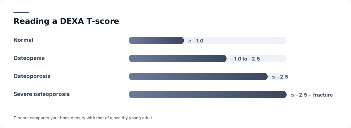
T-score compares your bone density with the average peak density of a healthy young adult of the same sex, expressed in standard deviations. It is the number used to diagnose osteoporosis in postmenopausal women and in men aged 50 and above.
- −1.0 or above: normal
- Between −1.0 and −2.5: low bone mass, or osteopenia
- −2.5 or below: osteoporosis
- −2.5 or below with a fragility fracture: severe, or established, osteoporosis
Z-score compares you with people of your own age, sex and body size. It is the appropriate measure in premenopausal women, men under 50 and children. A Z-score of −2.0 or below is described as "below the expected range for age" and should prompt a search for a secondary cause.
Osteoporosis can be diagnosed without a scan
A hip fracture from a fall from standing height, or a vertebral fracture without major trauma, establishes clinical osteoporosis regardless of what the T-score says. Density is one measure of bone strength, not the whole of it.
Who should be screened
Current U.S. guidance recommends bone density screening for:
- All women aged 65 and older.
- Postmenopausal women under 65 whose estimated fracture risk, using a formal tool, equals or exceeds that of a 65-year-old white woman without additional risk factors.
- Anyone who has had a fragility fracture after age 50.
- Adults on long-term glucocorticoids, generally the equivalent of 5 mg prednisone daily for three months or more.
- Adults with conditions associated with bone loss: hyperparathyroidism, hyperthyroidism, coeliac disease and other malabsorption, chronic kidney or liver disease, rheumatoid arthritis, HIV, anorexia nervosa, or after bariatric surgery or organ transplant.
- Men aged 70 and over, per several specialty organisations, and men 50 to 69 with risk factors. Evidence in men is thinner than in women, and screening rates are far lower than the fracture burden justifies.
The FRAX tool
FRAX estimates ten-year probability of a major osteoporotic fracture and of hip fracture, combining age, sex, weight, height, prior fracture, parental hip fracture, smoking, glucocorticoid use, rheumatoid arthritis, secondary osteoporosis and alcohol intake — with or without a femoral neck density value. In the United States, treatment is commonly considered when ten-year hip fracture risk reaches 3% or major osteoporotic fracture risk reaches 20%.
What raises risk
Fixed factors: older age, female sex, a parent who fractured a hip, small body frame, and being of white or Asian ancestry, which is associated with higher measured rates although osteoporosis occurs in all groups and is underdiagnosed in Black and Hispanic Americans.
Modifiable factors: smoking, heavy alcohol use, physical inactivity, low body weight, low calcium and vitamin D intake, and repeated falls.
Medications: long-term glucocorticoids are the leading drug cause. Others include some anticonvulsants, aromatase inhibitors, androgen deprivation therapy, long-term proton pump inhibitors, certain diabetes medications and depot medroxyprogesterone acetate.
Conditions: untreated hyperthyroidism, hyperparathyroidism, early menopause, hypogonadism in men, malabsorption and chronic inflammatory disease.
What happens after an abnormal result
First, a search for secondary causes. Blood tests commonly include calcium, phosphate, alkaline phosphatase, kidney function, 25-hydroxyvitamin D, TSH, and in men testosterone; parathyroid hormone and coeliac serology are added when indicated.
Then, treatment. The foundations apply to everyone:
- Calcium, ideally from food — dairy, fortified plant milks, tinned fish with bones, leafy greens, tofu set with calcium. Around 1,000–1,200 mg daily depending on age and sex.
- Vitamin D sufficient to support absorption, typically 800–1,000 IU daily in older adults.
- Weight-bearing and resistance exercise. Walking, stair climbing, dancing and progressive resistance training all stimulate bone. Balance work such as tai chi reduces falls, which matters at least as much as density.
- Fall prevention. Reviewing sedating medications, correcting vision, improving home lighting, removing loose rugs and installing grab bars prevents more fractures than most people realise.
- Stopping smoking and moderating alcohol.
Medication is added when risk warrants it. Bisphosphonates are usually first line. Denosumab, an injectable given twice yearly, is an alternative — and must not be stopped abruptly without transitioning to another agent, because rapid rebound bone loss and vertebral fractures can follow. Anabolic agents that build bone, including teriparatide, abaloparatide and romosozumab, are used in severe or very high-risk osteoporosis, typically followed by an antiresorptive drug to hold the gains. In some postmenopausal women, hormone therapy or a selective oestrogen receptor modulator is appropriate.
The treatment gap
One of the largest failures in this field is that most older adults who break a hip or wrist from a fall are never evaluated or treated for osteoporosis afterwards — despite that fracture being the strongest single predictor of the next one. If you or a relative has had a fragility fracture, ask explicitly about bone density testing and treatment. Many hospitals run fracture liaison services designed for exactly this.
How often to repeat the scan
Intervals depend on the initial result. Normal density or mild osteopenia in an older woman may not need repeating for ten to fifteen years, since progression is slow. Moderate osteopenia is often rechecked in three to five years, and advanced osteopenia or treated osteoporosis every one to two years. Comparisons are most reliable on the same machine, so continuity of facility matters more here than in most imaging.
The bottom line
A DEXA scan is quick, cheap, painless and one of the few tests that can identify a serious condition roughly a decade before it causes harm. The barriers are not technical — they are that women under 65 rarely have their risk formally assessed, that men are barely screened at all, and that fractures which should trigger evaluation routinely do not. Ask for the assessment, and ask again after any fracture.